Success Story
When reduced motility leads to disease
Article
Primary Ciliary Dyskinesia (PCD) is a rare genetic disorder affecting about one in 7500 people and is usually diagnosed in childhood. It is caused by dysfunctional motile cilia, which are tiny, hairlike projections that extend from the surface of certain cells in our body. Cilia can move and perform vital functions in various organs. For example, in the lungs they help to remove mucus and microbes in order to prevent infection. Cilia on sperm cells propel them forward. Similarly, in the female reproductive system, cells carrying cilia can transport the egg towards the uterus. And, in early embryonic development, these organelles are responsible for establishing a left-right body axis.
Cilia that do not function properly can cause a variety of symptoms, the most obvious of which are chronic respiratory infections of the lungs, sinuses or ears. However, a person with PCD suffering from respiratory symptoms may also have fertility issues, and in some persons, the internal organs are arranged mirror-imaged, with the heart lying on the right side. This is because the cilia in different organs are composed of the same proteins.
High resolution images of cilia
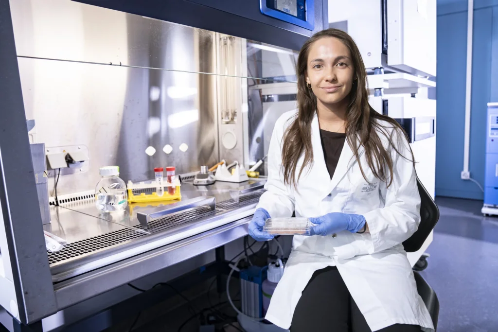
The overarching goal of the group led by Takashi Ishikawa at Paul Scherrer Institute (PSI) is to understand the cause of this disease and improve early diagnosis. Funded by PHRT, doctoral student Charlotte Ceuninck van Capelle applied high-resolution cryo-electron microscopy to PCD with the goal of molecular diagnostics.
To date, there is no available cure for PCD. There is also not one single test that can diagnose the disease. Cilia are composed of more than 300 different proteins. So far, the genes of around 60 of these proteins have been identified and can be tested for. “If PCD is suspected, the patient is usually referred to a specialized center where a biopsy is taken from the nose”, says de Ceuninck van Capelle. In Switzerland, this is done at the Inselspital in Bern, where, in addition to genetic testing, traditional electron microscopy and high-speed video microscopy are performed. “This provides information about whether the cilia are present, their structure, if they are beating properly, and if they are functioning correctly.”
However, in 20 to 30 percent of cases where the symptoms are very highly indicative of PCD, the genetic or structural causes cannot be found using genetic testing and electron microscopy. This is where the research project at PSI comes in. Using cryo-electron tomography, which involves freezing the sample at minus 180°C degrees, thus keeping it in its native state, the scientists can obtain high resolution images of the cilia. Taking pictures at different angles and then computing them into a 3d structure, provides far more structural information than conventional electron microscopy.
In addition to the high-resolution cryo-electron tomography, de Ceuninck van Capelle also performs mass spectrometry measurements. “This gives us a rough idea of which proteins are present in the sample”, she explains. “By combining these two techniques, we have an idea of both the structure of the cilia and their protein composition in any given patient with PCD”, she explains. This could shed light on subtypes that have previously remained hidden using conventional diagnostic methods.
So far, the group at PSI has studied over a dozen patients with PCD. In a recently published article, they report on three patients carrying genetic defects in the same gene and show that the precise location of the mutation can profoundly alter the internal architecture of human cilia. “So far, it is not very well known how different mutations in the same gene affect disease severity”, says Charlotte de Ceuninck van Capelle.
As cilia are composed of 300 different proteins, any of these proteins could potentially be involved in the disease. However, it appears that some proteins express the dysfunction more than others. In this study Charlotte de Ceuninck van Capelle focused on patients with defects in the most commonly affected motor protein.
Detecting previously unknown proteins
Recently, Inselspital Bern approached the PSI group to examine the structure of cilia in patients showing symptoms but testing negative in the genetic tests. “Using the cryo-electron tomography may help us to identify previously unknown genes involved in PCD”, she says. “In particular, this could help us detect small proteins that have not yet been identified using conventional electron microscopy.”
In addition to defects in the proteins that actually make up the cilia themselves, there may also be problems associated with cytoplasmic proteins known as chaperones, which help to transport or assemble the ciliary proteins. “Importantly, these are cytoplasmic, not ciliary proteins that may be defect”, explains Takashi Ishikawa. In this case, although the ciliary components are all expressed, they do not assemble correctly, thus resulting in dysfunctional cilia. Currently, these cases are sometimes missed by genetic testing.
This is why combining cryo-electron tomography with mass spectrometry is so essential. “Mass spectrometry will tell you whether a protein is present or not. But the cryo-electron tomography will tell you whether the protein has been transported and assembled correctly, says Takashi Ishikawa. This allows us to test several stages of cilia formation, adding new insight into the disease process.” The ultimate aim of the PSI scientists is to improve diagnosis. But before they can do so, they need a better understanding of the underlying biological processes. Cryo-electron tomography allows them to see the 3D structure of the protein and that is something new.
“With our research, we will hopefully be able to diagnose the disease earlier and develop targeted treatments that may halt or slow the progression of lung damage, hearing loss, and fertility disorders,” says Charlotte de Ceuninck van Capelle. And, thanks to PHRT, this endeavour has made quite some progress.